CBCT Cone BeamMansfield, TX
Dental cone beam computed tomography, or CBCT cone beam, is a new type of X-ray equipment that uses a 3D cone beam and dental scans to provide dentists with a detailed, three-dimensional image of the mouth. Furthermore, patients do not have to suffer any pain or discomfort during the procedure. A CBCT cone beam can bring about detailed photos of the bones, nerve pathways, soft tissues, and teeth with just a single scan.
CBCT cone beam is available at Toni Carr, DDS Inspiring Smiles in Mansfield and the surrounding area. This advance in dental technology may help save your teeth. Call us today at (682) 999-3696 to schedule an appointment or learn more about our services.
Cone Beam Computed Tomography
Inspiring Smiles is extremely proud to be able to offer our patients Cone Beam Computed Tomography (CBCT) diagnostic scanning using the Axeos, from Sirona, which is one of the most advanced dental scanning devices available.
What is CBCT?
In layman's terms, CBCT is a compact, faster and safer version of the regular CT. Through the use of a cone shaped X-ray beam, the size of the scanner, radiation dosage and time needed for scanning are all dramatically reduced.
The Axeos CBCT scanner fits compactly into our office structure and is easily accessible by patients. The time needed for a full scan is typically under 20 seconds and the radiation dosage is up to a hundred times less than that of a regular CT scanner.
- Visualize internal anatomy that cannot be diagnosed externally
- Plan treatment and surgery, including Implants
- Prepare necessary aids
- Assess risk and possible pathology and disease
- Analyze the position and orientation of critical structures, like nerves, teeth roots, previous implants, the sinuses and nose, and airway passages
What are the CBCT Scans used to diagnose and treat?
- Oral surgery
- Implant planning
- Orthodontic planning & implant anchorage
- Cephalometric analysis
- TMJ analysis
- Airway study (sleep apnea)
- Jaw tumors
- Impacted teeth
- Periodontal diseases
- Endodontic anomalies
Why a CBCT scanner rather than a regular Medical CT scanner?
- X-ray Radiation exposure to the patient is up 10 times less than a regular CT scanner.
- Much faster scan time. Scan on a CBCT takes between 10-40 sec, while on a regular CT scanner it takes a few minutes.
- Cheaper, the average price of a CBCT scan could be up to 50% less than a regular MDCT scan.
- What are the benefits versus risk?
Benefits
- Unlike regular X-rays, CT scans can discriminate between many types of tissue including bone, teeth, nerves and soft tissue.
- CT scans are noninvasive and can eliminate the need for exploratory surgery in some cases.
- CT can identify the effects of conditions such as infection and tumors.
- A cost-effective tool for imaging a wide range of clinical problems.
Risks
In a dosimetry study conducted by Dr. Ludlow (2007) of the University of South Carolina the Sirona Axeos has the lowest radiation dose of any cone beam machine on the market. One scan is compared to about 8 days of normal background radiation. This doesn’t even mean 8 days exposed in the sun but only 8 days of normal radiation by simply living on the planet earth. As with all imaging modalities that use ionizing radiation, the use of CBCT does involve a consideration of risk to the patient. However, it has the benefit of providing useful information needed to assist in making a diagnosis and/or in facilitating treatment. When your doctor refers you for an X-ray examination, he has made the determination that the benefit outweighs the risk. Of course, it is ultimately up to you to decide whether to undergo the examination. Bear in mind that the risk of most X-ray examinations are much less than other risks we commonly accept in daily life.
Understanding CBCT Cone Beam
A CBCT cone beam is a special type of X-ray machine utilized when regular dental or facial X-rays are insufficient. CBCT cone beams generate 3D images of dental structures, soft tissues, nerve paths, and bone in the craniofacial region in a single scan using a special kind of technology. As such, CBCT cone beam images allow the dentist to plan for more precise treatment.
While the CBCT cone beam is not the same as conventional CT, it can still be used to produce images similar to the ones made by traditional CT imaging. With a CBCT cone beam, an X-ray beam in the shape of a cone moves around the patient to produce multiple images, also known as views. Both CT scans and CBCT cone beam produce high-quality images, though CBCT cone beam utilizes a much smaller machine.
"A CBCT cone beam is a special type of X-ray machine utilized when regular dental or facial X-rays are insufficient."
How CBCT Cone Beam Works
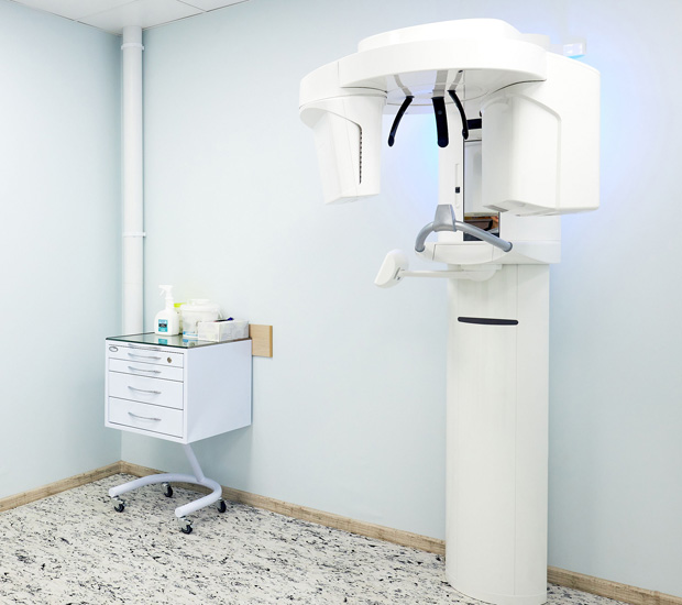
CBCT cone beam scanners are square-shaped machines, including an upright chair for patients to sit or a moveable table for patients to lie down during the examination. Scanners with a chair have a rotating C-arm, a device that intensifies X-ray images and contains an X-ray source and detector. Scanners with a table include a rotating gantry.
The C-arm or gantry will rotate around the head in a single 360-degree rotation, capturing multiple images from different angles during the examination. These images will then be reconstructed to create a single 3D image. The X-ray source and detector will be mounted on opposing sides of the revolving C-arm or gantry. They will rotate in unison, generating anywhere from 150 to 200 high-resolution 2D images. These are then digitally combined to form a 3D image, providing the dentist with valuable information about the patient's oral and craniofacial health.
"CBCT cone beam scanners are square-shaped machines, including an upright chair for patients to sit or a moveable table for patients to lie down during the examination."
CBCT Cone Beam Results
CBCT cone beam scanners bring forth far more detailed images than traditional CT scanners. Consequently, as previously established, they are particularly useful when traditional X-rays are insufficient to bring about the needed treatment information. For example, CBCT cone beams can evaluate the bony structures of the face, dentition, diseases of the jaw, nasal cavity, and sinuses.
CBCT cone beam technology can also be useful in diagnosing oral cancers and cysts and managing impacted teeth. According to one study, CBCT cone beam technology was found to have contributed significantly to the planning and successful surgical management of dentigerous cysts and associated impacted teeth. In addition, as cone beams take wide-range photos, they can capture in-depth areas that traditional scanners may miss.
"CBCT cone beam scanners bring forth far more detailed images than traditional CT scanners."
After the CBCT Cone Beam Procedure
Once the CBCT cone beam procedure has been completed, the dentist will have a clear, 3D image of the teeth, mouth, jaw, neck, ears, nose, and throat. It is possible to review this image immediately and get the best possible view of any area with features such as zooming and rotation. In addition, this is a painless procedure with no recovery time, and the patient will be able to return to their everyday activities immediately.
The dentist will also write a report of the scan results, and the patient will be able to see their images. Finally, the dentist and patient will go over the treatment plan together, during which time the patient can discuss their preferences for the treatment process and aftercare.
"Once the CBCT cone beam procedure has been completed, the dentist will have a clear, 3D image of the teeth, mouth, jaw, neck, ears, nose, and throat."
Questions Answered on This Page
Q. How do CBCT cone beam scanners work?
Q. What is the difference between CBCT cone beam scanners and traditional CT scanners?
Frequently Asked Questions
Q. Will my insurance cover CBCT cone beam scans?
A. Some insurance companies may cover CBCT cone beam scans. However, this is not a guarantee, as all plans are different. Your best bet is to contact your insurance provider directly. We can help you figure out what is covered in your plan.
Q. What kind of image is produced by a CBCT cone beam scan?
A. CBCT cone beam scanners take detailed images of the entire head. As teeth positioning affects the entire head, these images allow us to see the underlying bone structure and jawline. With this information, we can create the most accurate treatments possible. CBCT cone beam scanners are particularly beneficial for procedures around delicate areas. They are also useful for planning correctional procedures.
Q. How long does a CBCT cone beam scan take?
A. These scans are typically quite brief. For example, a CBCT cone beam scan only needs a singular rotation around the head. Once you are positioned, the scan itself will usually be over in less than 30 seconds.
Q. How long have dentists been using CBCT cone beam technology?
A. According to the Food and Drug Administration (FDA), dentists have been using CBCT cone beam technology for over twenty years. Furthermore, dental scans are becoming increasingly common, thanks to their helpfulness in procedure planning and diagnosing complex conditions.
Q. How much radiation does a CBCT cone beam scan emit?
A. Though CBCT cone beam scans are still considered computed tomography (CT) scans, they emit considerably less radiation than conventional CT scans. However, it does emit more radiation than a traditional X-ray. Therefore, we will only recommend a CBCT cone beam scan if we judge the benefits greater than the risks.
Q. How should I prepare for a CBCT cone beam scan?
A. There is very little preparation necessary for a CBCT cone beam scan. Make sure to remove all loose or metallic objects before the exam. Stay still and do not swallow or talk during the scan.
Definitions
- American Dental Association (ADA)
- The oldest and largest professional organization that represents dentists in the United States succeed in the advancement of public health through advocacy, education, and research.
- CBCT Cone Beam
- A special type of X-ray equipment used in place of regular dental or facial X-rays.
- Craniofacial Region
- The anatomical area that includes the skull and face, which encompass bones, muscles, and blood vessels.
- CT scan
- A diagnostic tool using a series of X-rays and a computer to detect diseases and injuries.
- Cysts
- An abnormal membranous sac or cavity containing fluid.
- Dental Eemergency
- A dental emergency is an acute condition affecting teeth and the surrounding tissue caused by trauma or infection that usually involves a lot of pain and must be dealt with immediately.
- Fractured Cusp
- A fractured cusp happens when a piece of the tooth’s chewing surface breaks off but does not affect the pulp chamber.
- Oral Cancer
- Any cancer originating in or around the mouth.
- Radiation
- The emission or transmission of energy in the form of waves or particles.
- X-Ray
- A digital image of the internal composition of a part of the body.
Call Us Today
CBCT cone beam can provide us with the necessary information to personalize an effective treatment plan for you. We at Toni Carr, DDS Inspiring Smiles in Mansfield, can help. Call us today at (682) 999-3696 to schedule an appointment or learn more about our services.
Toni Carr, DDS Inspiring Smiles is located at 1900 Matlock Rd. Suite 600 Mansfield, TX 76063.
Related Posts
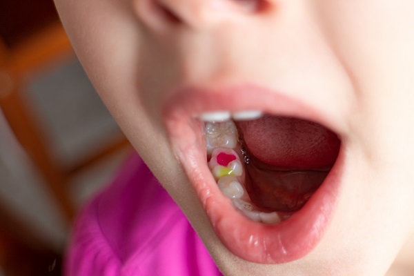
How A Tooth Filling Is Done
tooth fillings offer a simple approach with little to no pain. This dental restoration treatment has long been the standard for restoring and rebuilding teeth damaged by cavities, injury, or minor imperfections such as chips or cracks. When considering different dental restoration options, it is a good idea to get familiar with how each procedure…
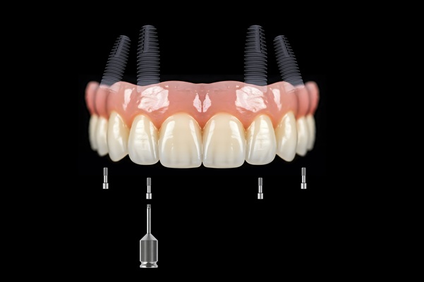
How To Care For Your All-on-4 Implants
If you have recently had All-on-4® implants, you may be wondering how to care for them. It is important to know the best way to keep your implants looking their best so they can last for many years to come. Here are the best ways to take care of your new smile.All-on-4 dental implants are…
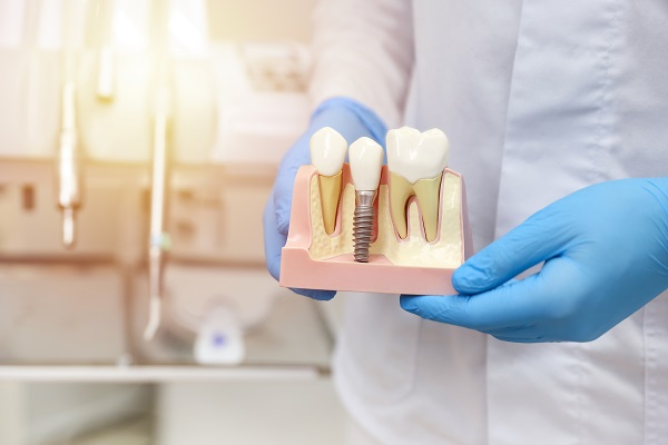
A Step-by-Step Guide To Implant Restoration
Implant restorations are a highly effective solution for replacing missing teeth, offering patients a durable, natural-looking option that restores functionality and aesthetics. They involve a process that combines the surgical placement of a dental implant with the creation and attachment of a prosthetic crown or other dental appliance. Take a look at this step-by-step guide…

Holistic Dentistry Near Me: Preparing For Your Appointment
If you are searching online for “holistic dentistry near me,” you are likely looking for a natural approach to dental care. Holistic dentistry prioritizes safe dental treatments that promote a healthy smile without compromising the body’s balance. Focusing on prevention and whole-body health offers a thoughtful alternative to traditional dental care. Making the most out…

What A General Dentist Looks For In Your Dental Exam
It is common for people to be nervous about their upcoming dental exam with a general dentist. Rest easy because there is no reason to be anxious about checkups like these. However, seeing a dentist twice a year is very important to your oral and overall health and safety.Dentists do much more than just look…